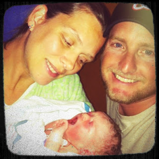
Ryleigh was born on 7/24, that post you have. I was discharged from the hospital the next day (good behavior and fast recovery).
Ryleigh was taken to the NICU the moment she was born (7/24/12 - 7/30/12). This was not because she had issues, but because her doctors played it safe, she was then hooked up to monitors and IV’s. She spent 7 days and 6 nights there learning all the wonderful things normal babies do, eating (small amounts to test her gut), filling her diaper, getting bathes…etc. The eating part was the best part because she learned how to develop those sucking skills so after surgery she can start right away and not risk losing weight. She also took to the paci right away which was also great! We got to hold her and love on her while she awaited surgery. It felt as normal as it could, being we were in a NICU ward and she was hooked to 20 different things. At first, it was hard to see because no momma wants to see her baby that way – but trust me it got worse. I recall them trying to get a PICC line in because the IV’s kept failing, tiny veins I guess. I came in one day and she had an IV in her head! The next day they shaved half her head to get the PICC line in her head, yikes, so glad they were able to get one in her arm instead.
What is a PICC line anyway?
A PICC line is, by definition and per its acronym, a
peripherally inserted central catheter. It is long, slender, small, flexible
tube that is inserted into a peripheral vein, typically in the upper arm, and
advanced until the catheter tip terminates in a large vein in the chest near
the heart to obtain intravenous access. It is similar to other central lines as
it terminates into a large vessel near the heart. However, unlike other central
lines, its point of entry is from the periphery of the body the extremities.
And typically the upper arm is the area of choice.
A PICC line provides the best of both worlds concerning
venous access. Similar to a standard IV, it is inserted in the arm, and usually
in the upper arm under the benefits of ultrasound visualization. Also, PICCs
differ from peripheral IV access but similar to central lines in that a PICCs
termination point is centrally located in the body allowing for treatment that
could not be obtained from standard periphery IV access. In addition, PICC
insertions are less invasive, have decreased complication risk associated with
them, and remain for a much longer duration than other central or periphery
access devices.
Using ultrasound technology to visualize a deep, large
vessel in the upper arm, the PICC catheter is inserted by a specially trained
and certified PICC nurse specialist. Post insertion at the bedside, a chest
x-ray is obtained to confirm ideal placement. The entire procedure is done in
the patient’s room
decreasing discomfort, transportation, and loss of nursing care.
A PICC line may requested for a variety of treatment options
which include some of the following:
-Prolonged IV antibiotic treatment;
-IV access obtainable by less invasive and longer lasting
methods;
-Multiple accesses obtainable with one access line;
-TPN Nutrition;
-Chemotherapy;
-IV access related to physiological factors; and
-Home or sub-acute discharge for extended treatment.
PICCs are frequently used to obtain central venous access
for patients in acute care, home care and skilled nursing care. Since
complication risks are less with PICC lines, it is preferred over other forms
of central venous catheters. A PICC is not appropriate for all patients. Proper
selection to determine the appropriateness of this device is required.
The PICC may have single or multiple lumens. This depends on
how many intravenous therapies are needed. A PICC line can be used for antibiotics,
pain medicine, chemotherapy, nutrition, or for the drawing of blood samples.
PICCs can be inserted by radiologists, physician assistants or certified
registered nurses. They are inserted using ultrasound technology at the bedside
or ultrasound wit fluoroscopy. Chest radiographs are also used to confirm
placement of the PICC tip if it was not inserted using fluoroscopy.Ok so let’s fast forward to the day she went to surgery…


No comments:
Post a Comment